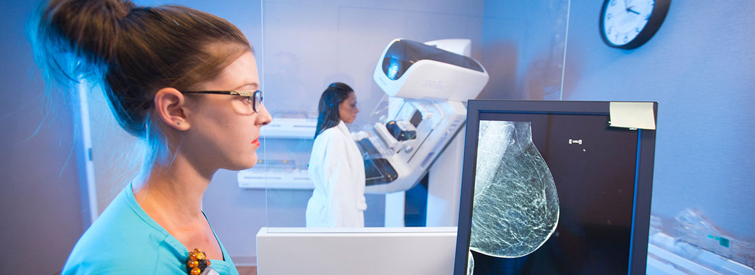
Digital mammography
Digital mammography allows doctors to see subtle differences in breast tissue that traditional film mammograms do not. In standard mammograms, images are recorded on film using an X-ray cassette. The film is then viewed by the radiologist using a "light box" and then stored in a jacket in the facility's archives.
With digital mammograms, the breast image is captured using a special electronic X-ray detector, which converts the image into a digital picture for review on a computer monitor. The digital mammogram is then stored on a computer.
The magnification, orientation, brightness and contrast of the image in digital mammography may be altered after the exam is completed to help the radiologist more clearly see certain areas.
That means more accurate diagnoses and fewer biopsies. Digital mammograms are faster – delivering less radiation exposure and giving faster feedback to patients.
Computer-assisted detection (CAD)
Using sophisticated computer software, CAD works essentially as a second opinion of the radiologist’s interpretation of a woman’s mammogram. The software marks even the smallest breast abnormalities on the mammogram image, so the radiologist can take another look and determine if further tests are required. The radiologist still makes the final interpretation of the mammogram.
Tomosynthesis (3D) mammography
Tomography (also known as 3D mammography) can be used for all women. However, women with dense breast tissue may benefit the most since dense breast tissue tends to overlap and can hide cancer.
Currently, 3D mammography is performed in combination with 2D mammography with same views, positioning and compression. A 3D mammogram is made by computer reconstruction of images obtained while the machine sweeps over the breast in an arc - sometimes in as little as 4 seconds for average size breasts. The images are taken at different angles so the computer can turn 2D into 3D. The radiologist can view them as moving pictures.
Benefits of 3D mammography:
Elimination of overlapping, compressed breast tissue improves our ability to find cancer earlier or find cancer that may be missed on 2D mammography alone.
Better visualization of fine details.
Greater accuracy determining size, shape and location of abnormalities.
Reduction in the number of call backs for additional mammograms - reduction of stress for women
Decrease in false positives (abnormality identified on the mammograph that is NOT cancer)
20-40% decrease in the mortality rate of breast cancer
The Radin Breast Care Center is proud to offer this exciting breakthrough technology as it will have a positive impact on breast cancer screening and diagnosis.
Follow-up diagnostics
Almost all women who get mammograms find they are in good health. But in cases where abnormalities are found, additional tests may be needed. The good news is about 80 percent of women who find abnormalities in their mammograms do not have cancer.
The Radin Breast Care Center offers technologies and services that allow your doctor to go beyond mammography and get a closer look at your health.
These technologies and services include:
Breast MRI
- is a test used to detect breast cancer and other abnormalities in the breast. Breast MRI is used when your doctor needs more information than a mammogram, ultrasound or clinical breast exam can provide.
Breast MRI captures multiple pictures of your breast and images are combined, using a computer, to generate detailed pictures needed to obtain images with high quality and clarity.
3D breast biopsy
- The Veranda technology provides the latest in imaging technologies for 3-dimensional breast biopsy. Paired with new 3D mammography, this biopsy system is able to detect 40% more invasive breast cancers than traditional mammography imaging alone. And, more than 80% of brease biopsies are normal.
Advantages of using this new breast biopsy technique include faster targeting with a shorter patient procedure time and fewer X-ray exposures resulting in a reduced X-ray dose.
Stereotactic core biopsy
- uses X-rays taken from multiple angles to pinpoint the center of irregular breast tissue. Taking a sample from the tissue core allows physicians to remove a small amount of tissue without the incisions, anesthesia and scarring associated with traditional biopsies.
Ultrasound core biopsy
- uses multiple images to pinpoint and sample abnormal breast tissue. There is less scarring, faster results and less pain than with traditional biopsies.
Fine needle aspiration
- is a technique that allows a biopsy of various bumps and lumps. It allows for enough tissue to be retrieved for microscopic analysis to make an accurate diagnosis.
Cyst aspiration
- uses a small needle to drain fluid from a cyst. Doing so can help your doctor find out if the lump you feel is a cyst or not.
Lymphedema therapy
- is now offered at the Radin Breast Care Center. Lymphedema is the swelling of the arm caused by an abnormal accumulation of protein rich fluid and can occur after breast cancer surgery, lymph node removal or radiation treatments.
This is a chronic condition which can worsen over time causing limited range of motion, repeated infection with possible hospitalization and a decrease in ability to care for oneself.
Symptoms include swelling, aching, tightness, heaviness and acute or chronic infections. Treatment consists of manual lymph drainage, application of compression bandages and garments, education for precautions and self management techniques. We offer one-on-one treatment sessions with a certified lymphedema therapist.
Personalized consultation with a team of specialists
- women tell us they want their doctors to coordinate better, so we give you the chance to meet together with the specialists who will treat you. The mix of specialists is tailored to each patient.
This multidisciplinary team of specialists meet to discuss a woman's coordinated treatment plan at the Radin Breast Care Center – which can be done in a faster timeframe and eliminate your need to wait to see these specialists at separate locations elsewhere.
Cancer care experience
- when test results show breast cancer is present, it is comforting to know we are affiliated with the California Cancer Center and the only cancer program in the area to earn “teaching hospital level” accreditation with commendation from the American College of Surgeons – an accreditation on par with university-based cancer programs at Cedars-Sinai Medical Center and UCLA Harbor Medical Center.
Support group: Luchadores contra cancer
Nuestro grupo de apoyo está aquí para ayudar a las personas que hablan Español, y que han sido diagnosticados con cáncer. Tambíen invitamos a los cuidadores qué participen en el grupo de apoyo.
Ofrecemos un lugar seguro para compartir sus experiencias, conectarse con otras personas afectadas por el cáncer y la oportunidad para aprender sobre su salud. Para más información, llame a Alva Reyes (559) 387-1872.
Cuando:
Segundo miércoles de cada mes
3-4 p.m.
Nurse coordinator
- the nurse coordinator at the Radin Breast Care Center makes sure you get all the care you need and that your experience with us is a good one. The nurse coordinator also serves as a knowledgeable resource for patients while coordinating care and providing education for each woman who comes through the center.
For more breast cancer information
Susan G. Komen Breast Cancer Foundation
We use cookies and other tools to optimize and enhance your experience on our website. View our Privacy Policy.